
I am a PhD researcher in the field of holography and an artist who is working in the emergent art form holography. I am not an anatomist, nor an anthropologist. I am describing a journey that leads to the creation of an artwork, and I am sure that there are many details that could be added.
I have been dreaming of making a hologram of the internal clitoris, or more correctly the bulbs-clitoral organ, with the surrounding anatomy since 2013 when I first saw a 3D model of it online. I have not been able to attribute the original source of this (widely shared) image.

For me personally, the first time I heard about the clitoris being anything else than the little thing in the upper part of the vulva was in 2008, reading the Swedish sex education/political magazine ‘Ottar’ at a friend’s house. I remember feeling quite angry that I, as a forty-year-old educated woman, had never heard of this “larger structure” before. There was no anatomically illustrative image accompanying this article.
https://www.ottar.se/artiklar/klitoris-story
I have met some resistance within academia to including a work on the theme in my practice-based arts research, but I have also received support as I insisted on standing by the idea. I have thought both once and twice about whether I want this piece to define me in my academic field of holography. But then I just had to do it! The work called me.
My experiences echo the interviewees in the ‘The clitoris coverup: why do we know so little‘?’ podcast, the Guardian of 31 October 2020.
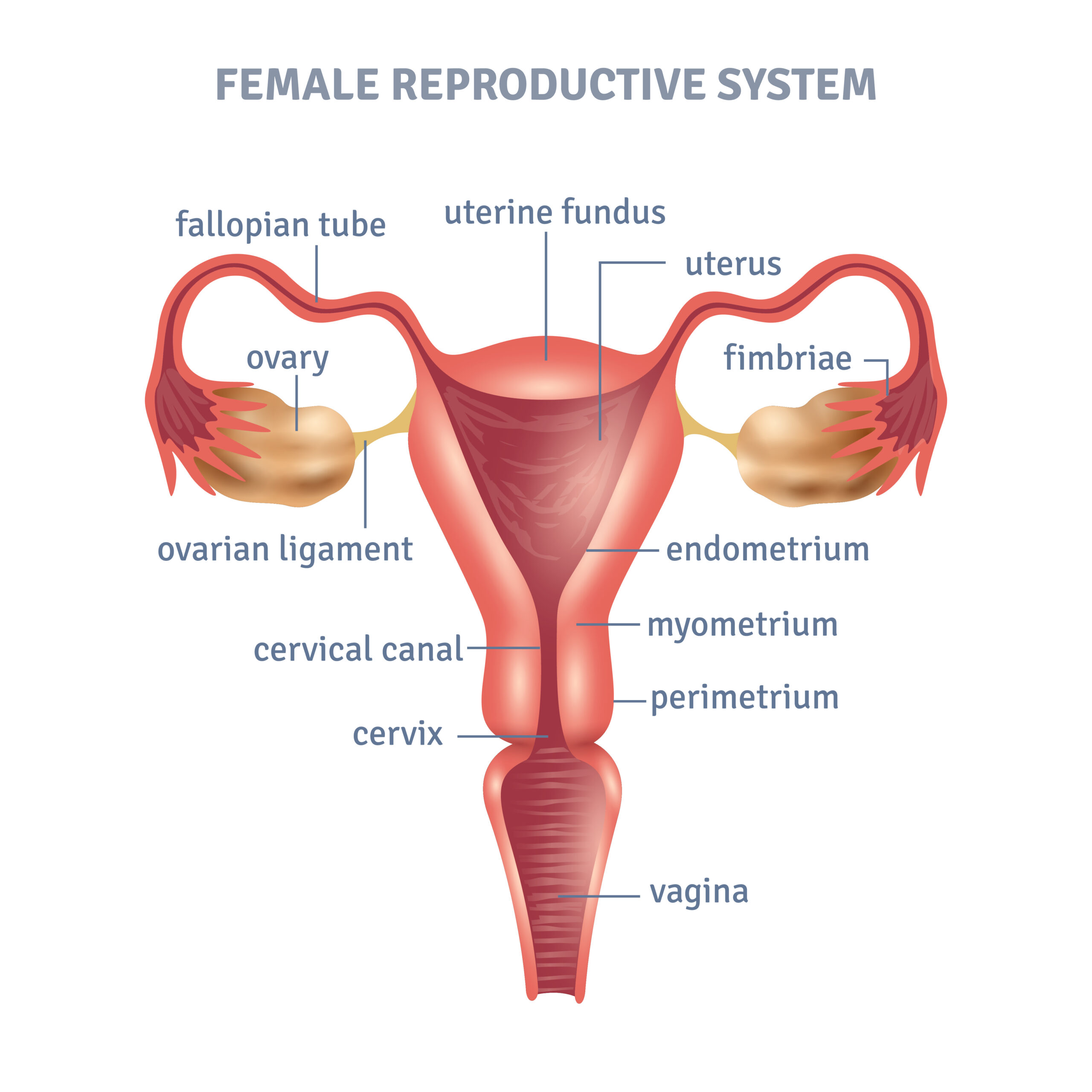
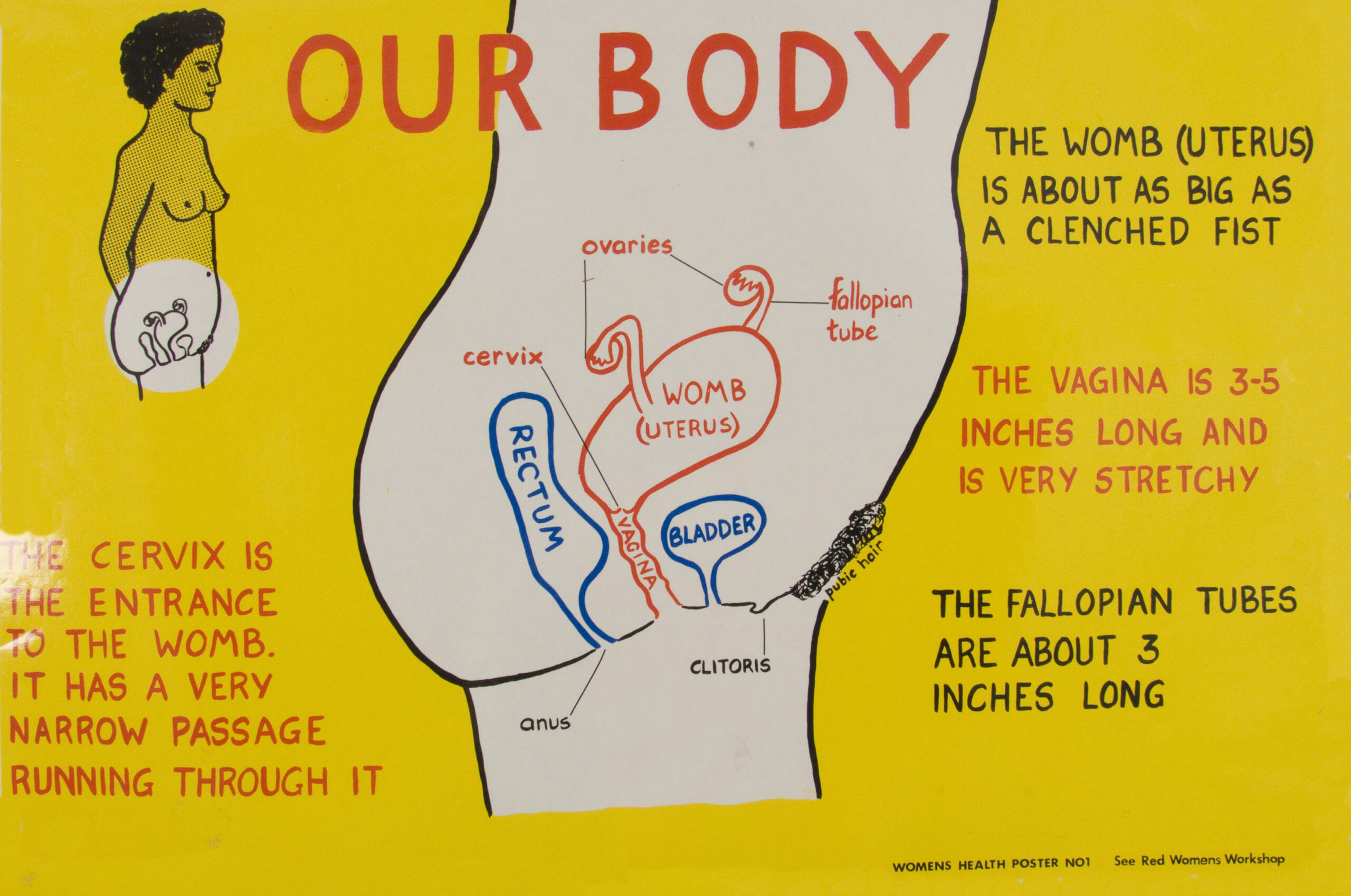
During my lifetime most representations of the female reproductive system looks something like the above image.
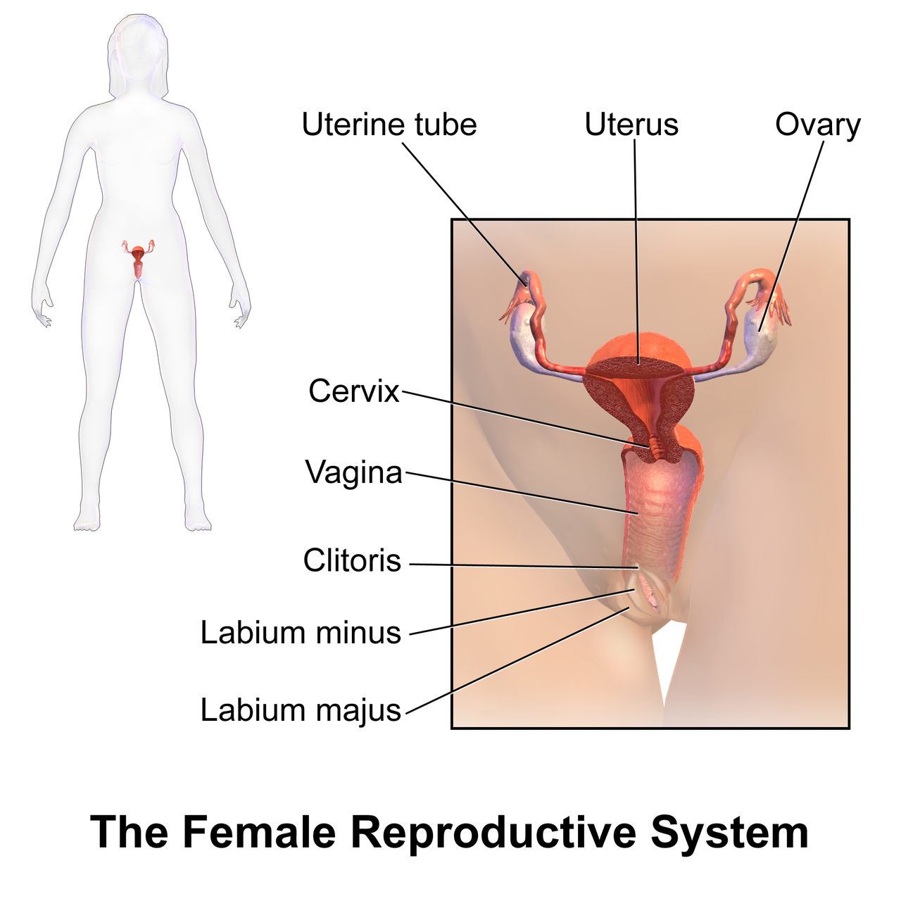
When the clitoris is included in the diagrams it was/is often portrayed as a very small anatomical feature.
This was/is also reflected in text, for example in Oxford English Dictionary, second edition (1989): “A homologue of the male penis, present, as a rudimentary organ, in the females of many of the higher vertebrata.”
And in the American Heritage Dictionary of the English Language, fifth edition (2021):
“A small erectile body situated at the anterior portion of the vulva and projecting between the branched extremities of the labia minora forming its prepuce and frenulum.”
It would be interesting to undertake a full investigation of how the definition of the clitoris has changed through the years. But this will suffice as an illustration.
However, the later “forgotten” larger structure of the clitoris was known to anatomists in the mid 1800s, as evident in this dissection illustration by Georg Ludwig Kobelt. One wonders why it subsequently disappeared for 170 years?

I want to celebrate the work of urologist Helen O’Connell (The Guardian article here) and researcher Odile Fillod (website in French here) who brought new research and anatomical understanding to a larger public in the current century.
Perhaps my work can be seen as sitting in a historical context of feminist educational art. To me it is a future iteration of this work ‘Our Body – Womens health poster no1’, produced by The See Red Women’s Workshop in the nineteen eighties. See Red Women’s Workshop Feminist Posters 1974 – 1990.
Some technical challenges for the contemporary holographer
Following the closure of the Zebra Imaging holography company 2017, there has not been any set up globally (!) that could expose my 3D digital model in a full-parallax format.
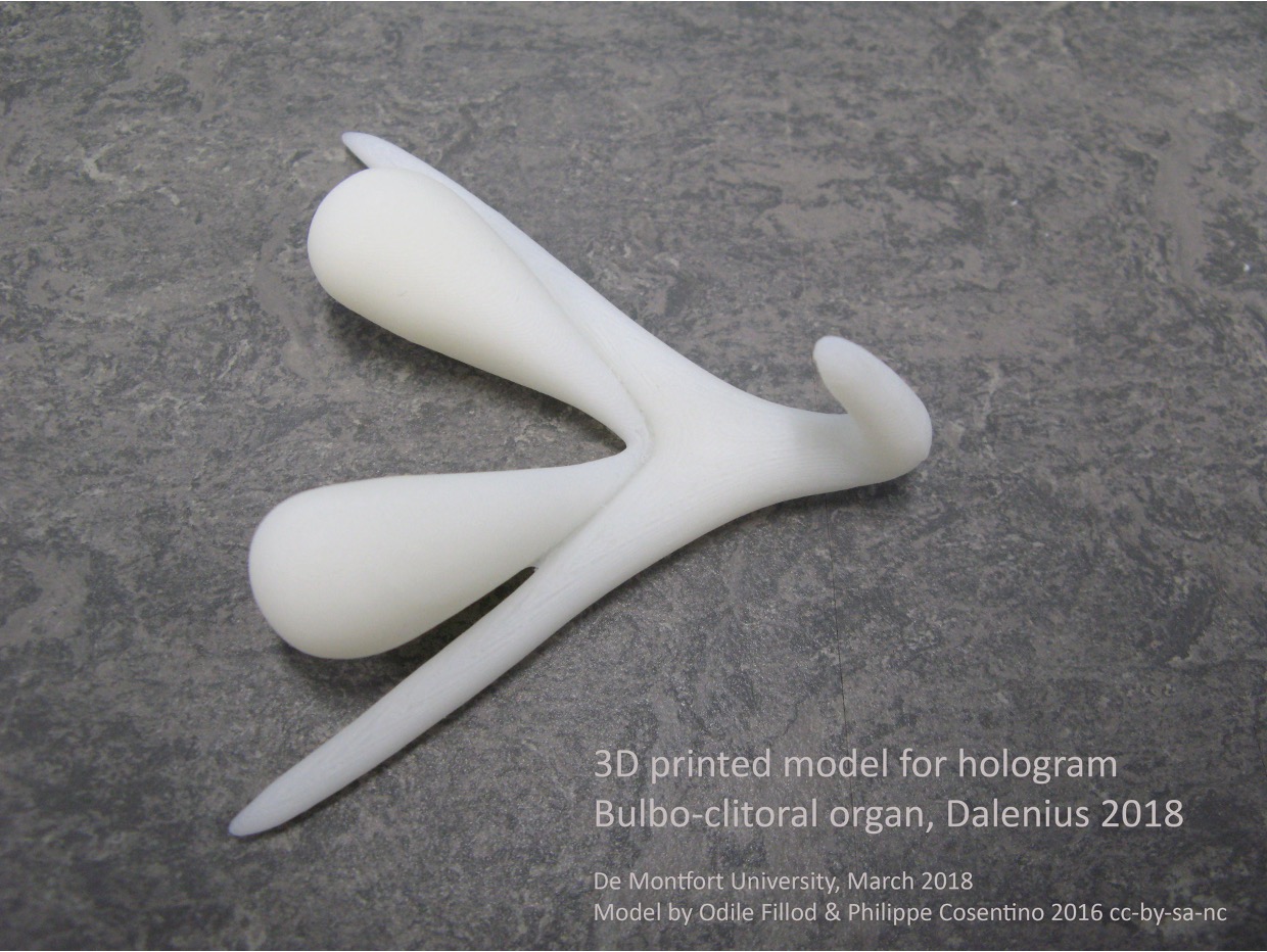
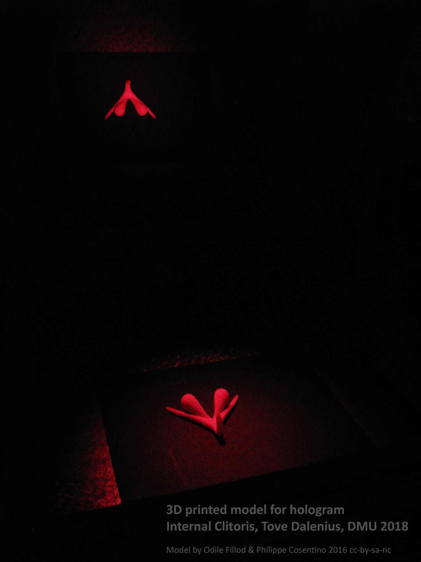
Although experimenting with analogue holography using a 3D printed physical model of the clitoris was exciting, I could not achieve my full vision for the piece in this format.
However, Yves Gentet has now developed the Chimera technology, which as well as offering a perspective field of view of 120° which provides full-parallax, uses 250 um (micrometers) holopixels. This ensures that the holographic image is smooth, and not “holopixelated”.
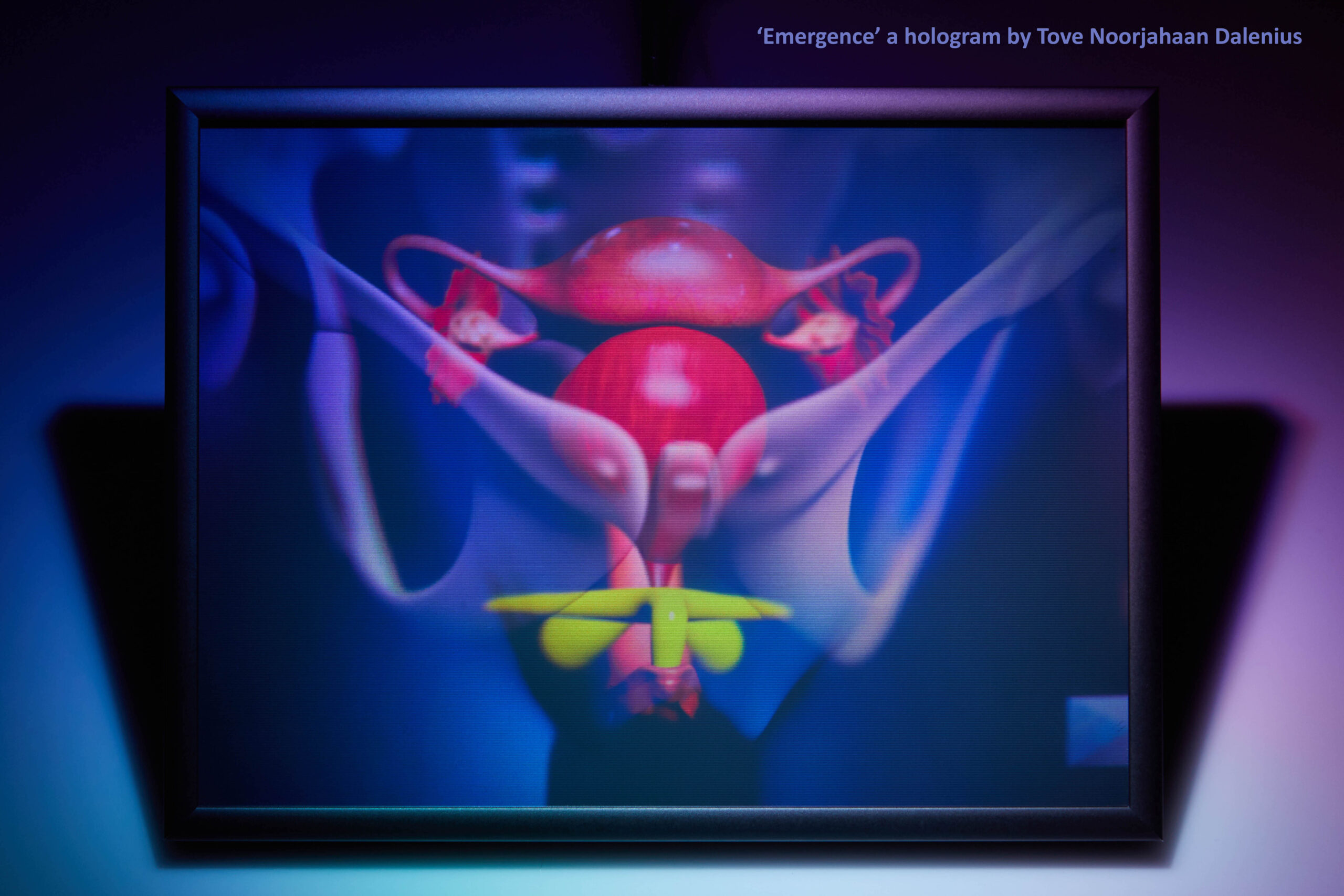
I used a model of Odile Fillod’s 3D printable model (http://carrefour-numerique.cite-sciences.fr/wiki/doku.php?id=projets:clitoris) and fitted it into a model of the female reproductive organ. Using a variety of 3D modelling programs such as Blender and 3D Studio Max, and the graphics editing program Photoshop.
Then came the whole process of turning it into a hologram – a 3D light sculpture.
As an artist, I aimed to compose the image within the three available dimensions and, in this piece, I attempted to avoid any “depth blur”. There was deliberation over the placement of digital objects, size, transparency, and colour. Even with years of experience, it is difficult to fully envisage how the completed hologram will appear, transformed from the 2D screen.
I am making a piece of art, and although the piece is both technical and educational, it is also spiritual to me. The space of artistic creativity has to be found and cherished. I wanted the hologram to be as anatomically correct as possible, but there is also an area in space where stars are born overlaying the uterus. I decided to include a platonic solid, an octahedron, with my signature in the work.
The work fills me with energy and peace when I see it. The Marian blue of the skin of the model reminds me of icons, perhaps a transparent veil? Inside there, in the uterus, stardust is assembled into new life. Consciousness is formed at some point in time. I stop and meditate in a silent room with the hologram. Why was the clitoris hidden and forgotten?
Why is even the most basic anatomy concealed from our knowledge? I want to encourage women to take ownership. Over technology, their bodies and art – but I hope that the work conveys this, without using too many words.
Dalenius, Tove Noorjahaan (2020)
Published January 2023
‘Emergence’
Hologram
15×20 cm
17 cm z-axis depth

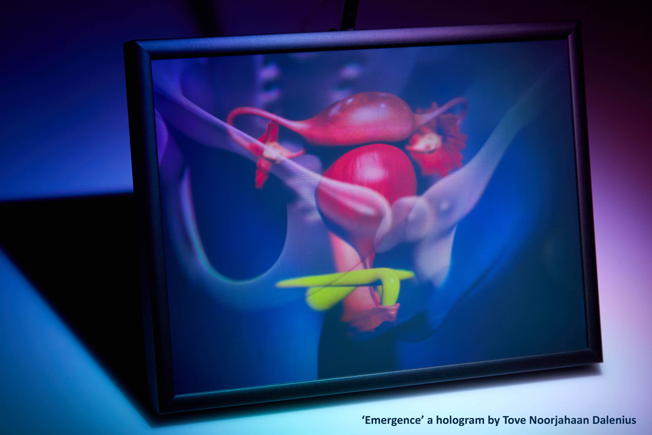
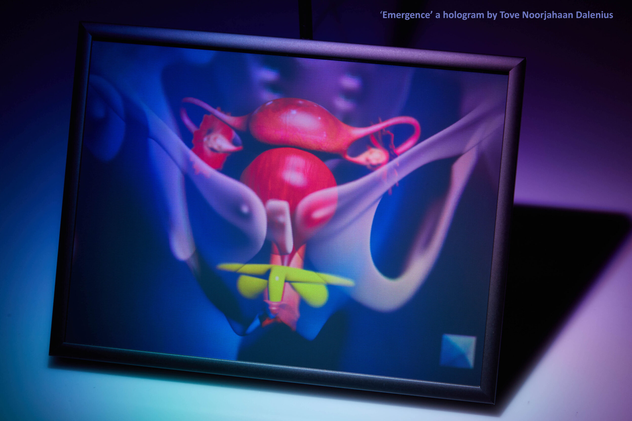
‘Emergence’ credits:
- Bulbo-clitoral organ model (in yellow) “Clitoris V2”: Odile Fillod & Philippe Cosentino
- Original image by NASA/ESA and Orsola De Marco (Macquarie University), 3D mapping by Tove N. Dalenius
- Stock 3D anatomy model
- CHIMERA holographic exposure Yves Gentet
- Holography composition Tove N Dalenius